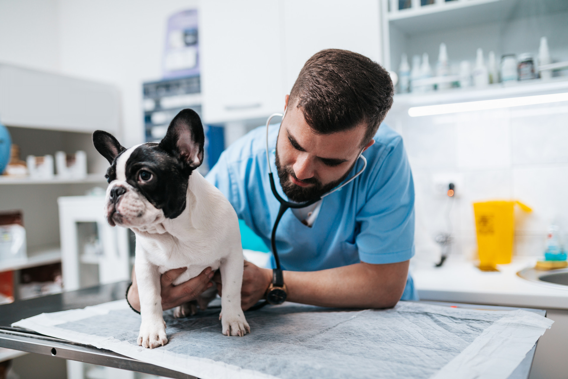
Early detection using diagnostic testing provides a complete picture of your pet’s health, improving outcomes, and providing baseline results.
As a pet owner, you might recognize when your pet is not feeling well. But it’s difficult to determine why.
Our staff is committed to helping you get a full picture of your pet’s health through laboratory and diagnostic testing.
Related Services
From preventative screenings to sophisticated diagnostics.
Blood & Lab Tests
Knowing when your pet is unwell is one of the most challenging parts of pet care. Annual blood and lab tests help determine a normal baseline for your pet's health. Monitoring these results over time allows us to diagnose problems early, increasing the likelihood of timely intervention and recovery so your pet can live their best life.
Ultrasound
Using sound waves, a vet ultrasound generates images of the inside of your pet's body, such as muscles, tendons, joints, organs, and blood vessels. Ultrasound images are often ordered by your vet alongside x-rays, to get a more complete picture of your pet’s health.
Tonometry
Tonometry is a non-invasive diagnostic test that measures the pressure inside your pet’s eyes. Increased pressure in your pet’s eyes can be dangerous and can lead to diseases like glaucoma and uveitis. Tonometry is an easy way to check the pressure of your pet's eyes without having to sedate them.
Electrocardiogram (EKG)
An EKG is a gentle test we use to listen to what's happening in your pet's heart. It can spot if their heart is too big, if there's a murmur, signs of heart failure, or if the beat is off rhythm. We also check with an EKG before any surgery to make sure their heart can handle the anesthesia. It's completely painless—your pet won't feel a thing! This test is a crucial step in making sure we take the best care of your pet's heart.
Cookies on this website are used to both support the function and performance of the site, and also for marketing purposes, including personalizing content and tailoring advertising to your interests. To manage marketing cookies on this website, please select the button that indicates your preferences. More information can be found in our privacy policy here.
We've upgraded our online store!
Ordering your pet's favorite food and medicine is now easier than ever.
Order Food & Meds
Quick & Easy Registration

Please use the phone number and email you currently use for hospital communications to link your account!
Linked Pet Records & Rx

Your pet's prescriptions and records will be waiting for you!
Pawsome
Savings!

AutoShip discounts, promotions on your favorite products and more!


