
We've upgraded our online store!
Ordering your pet's favorite food and medicine is now easier than ever.
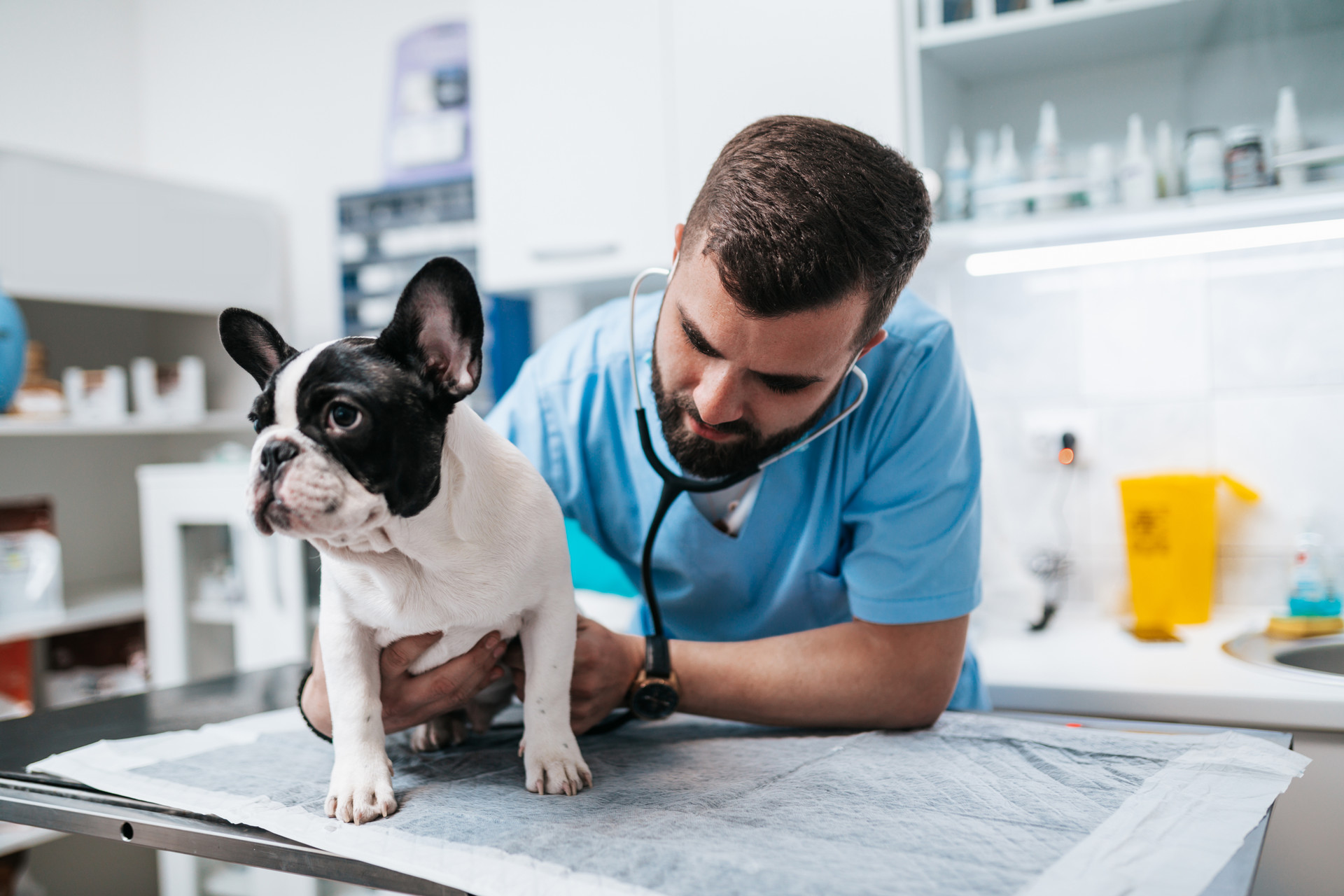
Order Food & Meds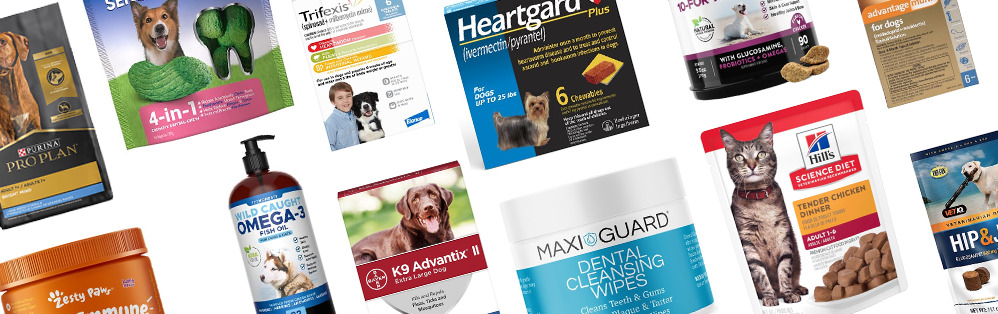
Quick & Easy Registration

Please use the phone number and email you currently use for hospital communications to link your account!
Linked Pet Records & Rx
Your pet's prescriptions and records will be waiting for you!
Pawsome
Savings!

AutoShip discounts, promotions on your favorite products and more!